
Build your model
We don’t believe in one-size-fits-all. Our models are designed to be flexible, modular, and tailored to your specific goals. Whether you need a single component or a complete solution, we can puzzle the pieces together to create exactly what works for you. Every project is built with precision, adaptability, and innovation—because your challenges deserve more than standard answers.


Applications


Explore our case studies
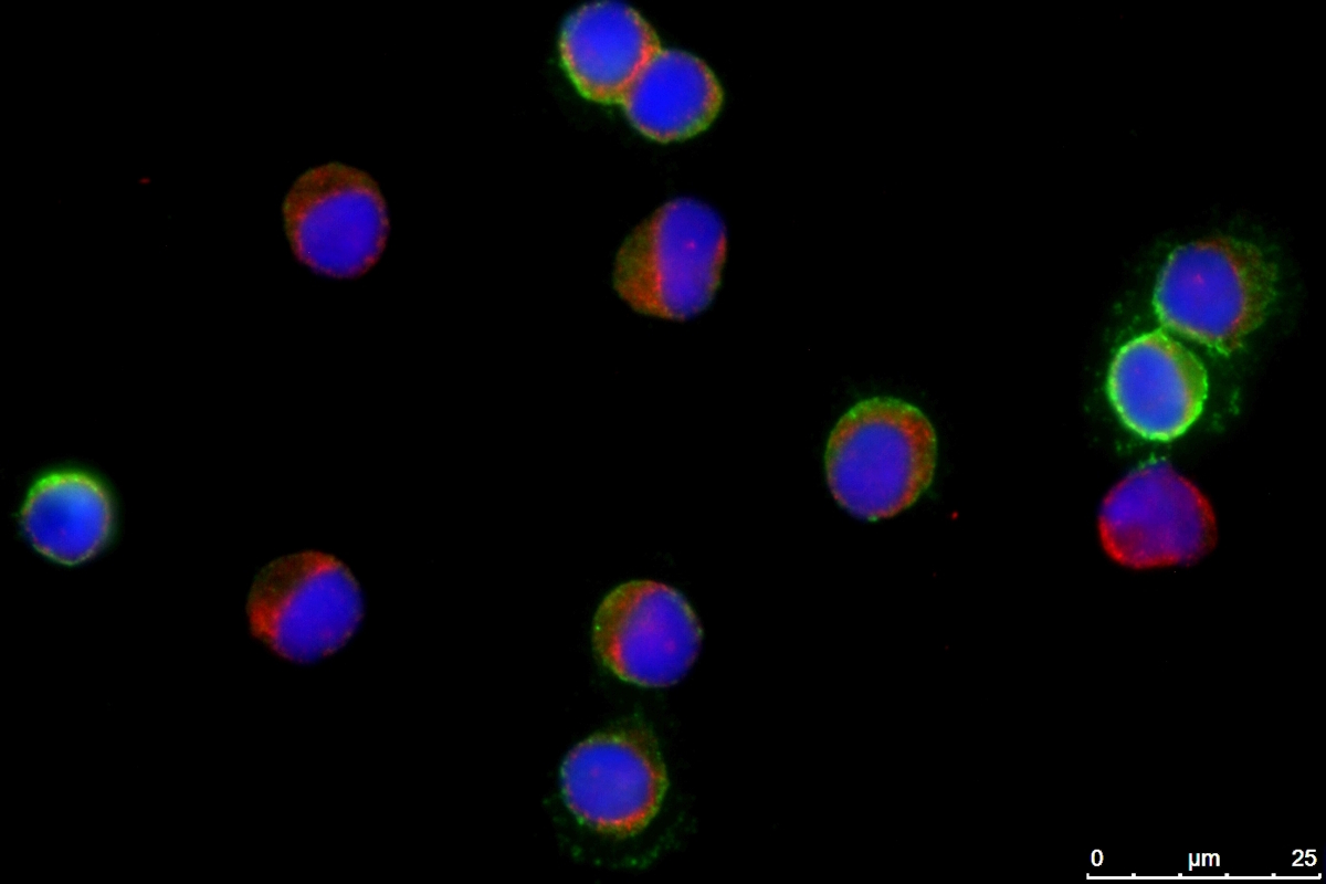
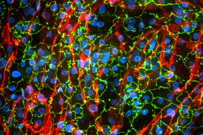
Healthy
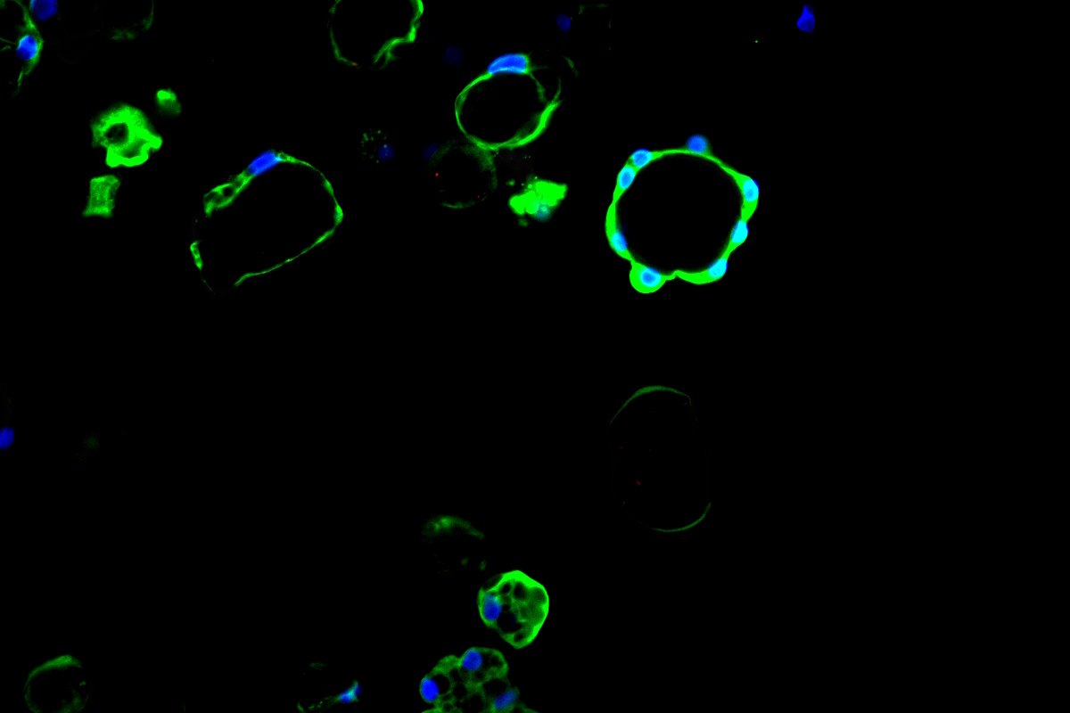
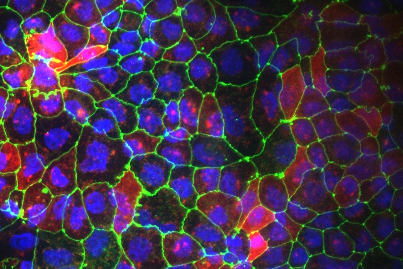
Diseased

AXLung-on-Chip
Learn more
AXGut-on-Chip
Learn more
AXKidney-on-Chip
Learn more
Interstitial Lung Disease (ILD)
Learn more
Inflammatory Bowel Disease (IBD) / Crohn's disease
Learn more

Chronic Obstructive Pulmonary Disease (COPD)
Learn more

Infection models
Learn more

Pulmonary Hypertension (PH)
Learn more
